Figure 3.
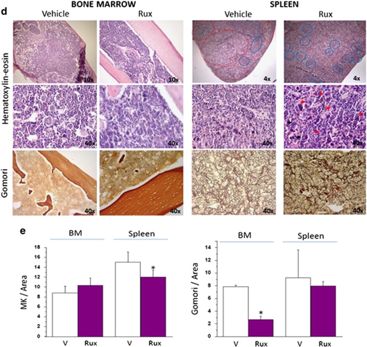
Treatment with Rux reduces JAK2 and restores the architecture of the spleen of Gata1low mice. (a, b) Determination of body weight, hematocrit (Htc) and platelet counts (Ptl) in female (a) and male (b) Gata1low mice treated for 2 weeks with either vehicle (V) or Rux, as indicated. For the group of males, determinations observed before treatment (pre) are also depicted. Results are presented as mean (±s.d.) with three mice per group and are compared with mean (±s.d.) observed in historic wild-type littermates, indicated by the horizontal boxes. *P<0.05 (by analysis of variance (ANOVA)) with respect to V. (c) Western blot analyses of JAK2 and STAT5 in spleen from Gata1low mice treated either with vehicle (V) or Rux (T) (each lane a separate animal). Quantification of results obtained with three to four mice per group is presented on the right. P-values were calculated by ANOVA. (d) Representative hematoxylin–eosin (top and middle panels on the left) and Gomori silver (bottom panels on the left) stains of bone marrow and spleen sections from Gata1low mice treated with either vehicle or Rux, as indicated. Note that the spleen of the Rux-treated mouse presents the typical dark red color of this organ with areas of white pulp (blue circles) appropriately located in germinal centers in the subcapsular area. By contrast, the spleen from vehicle-treated mouse has a disorganized morphology with excessive red pulp areas and paucity of white pulp areas localized mostly in the middle of the parenchyma. Original magnification × 4, × 10 and × 40, as indicated. Representative megakaryocytes are indicated by arrow. (e) Computer-assisted quantification of the frequency of megakaryocytes (MK) and of the intensity of Gomori silver staining of bone marrow (BM) and spleen from Gata1low mice treated with either vehicle or Rux (three mice per experimental group). *P<0.05 by ANOVA.


