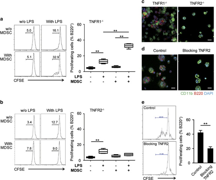
Figure 4.
TNFR2 is necessary for MDSC-mediated B-cell proliferation in vitro. (a, b) CFSE-labeled non-adherent spleen cells were cultured alone or with TNFR1−/− (a) or TNFR2−/− MDSCs (b) in the presence or absence of LPS (1 μg/ml). The B220+ cell division was assessed by flow cytometry for CFSE dilutions after 72 h. The left panels show representative histogram plots and percentages of the divided cells. The right panels summarize three independent experiments with triplicate determinations. *P<0.05 and **P<0.01, as determined with the Mann-Whitney test. (c, d) Non-adherent spleen cells were co-cultured with TNFR1−/− or TNFR2−/− MDSCs (c) or with wild-type MDSCs in the presence or absence of antibodies specifically blocking TNFR2 (100 ng/ml) (d) for 72 h. LPS (1 μg/ml) was added into all of the cultures in c and d. The adhesion of B220+ (red) cells and CD11b+ MDSCs (green) cells was visualized by cLSM. The nuclei were counterstained with DAPI; scale bars, 20 μm. (e) CFSE-labeled non-adherent spleen cells were cultured with MDSCs in the presence of LPS with or without TNFR2 blockade (100 ng/ml), and the B220+ cell division was assessed by flow cytometry as a CFSE dilution after 72 h. The left panels show representative histogram plots and percentages of the divided cells. The right panels indicate mean values±s.e.m. of the percentages of the divided B220+ cells from three independent experiments with triplicate determinations. *P<0.05 and **P<0.01, as determined with Student’s t-test. CFSE, carboxyfluorescein succinimidyl ester; cLSM, confocal laser scanning microscope; MDSCs, myeloid-derived suppressor cells; TNFR, tumor necrosis factor receptor.

