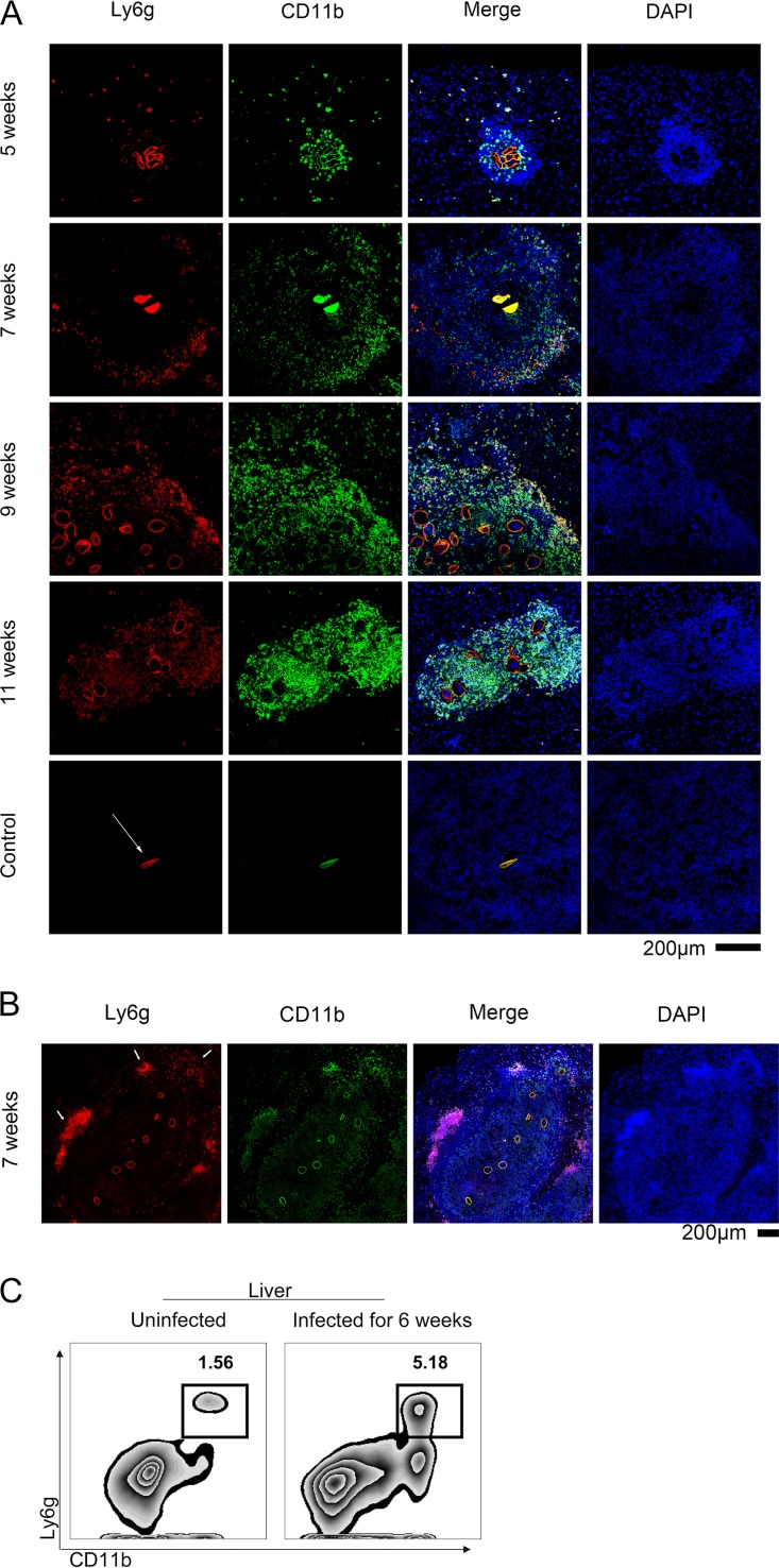
FIG 2.
Pathological changes in the liver caused by neutrophils. (A) Liver tissue samples were obtained from C57BL/6 mice infected with S. japonicum at the indicated weeks postinfection and stained with PE-conjugated-anti-Ly6G and FITC-conjugated anti-CD11b antibodies. Representative images are shown. The white arrows indicate the shell of the egg, which is always brightly colored in red (ly6G+) and green (CD11b+) but is invisible in the blue channel. Scale bar, 200 μm. (B) A large granuloma and several eggs were observed in the liver 7 weeks postinfection. The arrows indicate neutrophils surrounding the new eggs and older neutrophils around the edge of the granuloma. Scale bar, 200 μm. (C) The percentage of neutrophils in the liver was assessed by flow cytometry. All of the control group consisted of unstained slides from WT mice infected with S. japonicum.

