FIG 5.
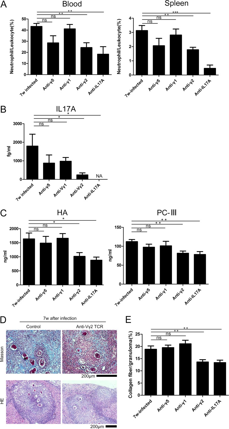
Depletion of Vγ2 T cells reduced the number of neutrophils in the blood and spleen. C57BL/6 mice were infected with S. japonicum (n = 5 mice/group), and then Vγ2 T cells were depleted using a monoclonal antibody 6 weeks postinfection. Liver, blood, and spleen samples were collected 8 days after depletion. (A) Summary graphs showing the percentages of neutrophils/CD45+ cells in the blood and spleen with and without depletion. (B) Summary graph showing the IL-17A level in serum with and without depletion. NA, not applicable. (C) Summary graph exhibiting the HA and PC-III levels in the serum with and without depletion. (D) Representative image of liver tissue stained with Masson's trichrome staining to measure fibrosis (blue). The image of the liver tissue stained with hematoxylin and eosin (HE) is shown. Scale bar, 200 μm. (E) Summary graph showing fibrosis as a percentage of the granuloma with or without depletion (number of granulomas observed in each group: 30, 28, 21, 44, and 30). ns, P > 0.05; *, P < 0.05; **, P < 0.01; ***, P < 0.001 (by Student's t test).

