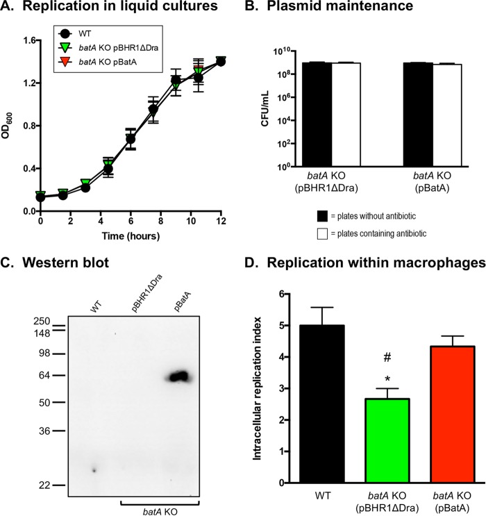
FIG 4.
In vitro replication rates and BatA production of B. mallei WT and recombinant strains. (A) Plate-grown bacteria were suspended in broth (without antibiotic supplementation) to an OD600 of 0.1. Following this, the bacteria were incubated at 37°C with shaking (200 rpm), and the optical densities of the cultures were measured from duplicate samples at the indicated time intervals. Strains were tested on 5 separate occasions. The error bars correspond to standard errors of the mean. The results of one representative experiment are shown. (B) At the endpoints of growth experiments, the liquid cultures were serially diluted and plated onto agar medium containing kanamycin (a selective marker encoded by plasmids pBHR1ΔDra and pBatA); duplicate aliquots were plated onto plain agar medium (no antibiotic added). The agar plates were incubated at 37°C for 48 h, and the CFU were counted to evaluate plasmid stability in recombinant strains. The error bars correspond to standard errors of the mean. The results of one representative experiment are shown. (C) Proteins were extracted from WT B. mallei ATCC 23344 bacteria and the batA KO mutant strain carrying the plasmids pBHR1ΔDra (control) and pBatA (specifying the WT batA gene) and analyzed by Western blotting with the monoclonal antibody BatA-MAb 1. Molecular mass markers are shown on the left in kilodaltons. (D) Plate-grown bacteria were suspended in PBS and used to infect 2 wells of duplicate tissue culture plates seeded with murine J774 macrophages (multiplicity of infection = 10:1). The infected cells were incubated for 1 h at 37°C to allow phagocytosis of the bacteria, washed, and treated with antibiotic for 2 h. Cells from one tissue culture plate were lysed, diluted, and plated onto agar medium to determine the number of bacteria phagocytized. The other tissue culture plate was incubated for an additional 7 h, after which the cells were washed, lysed, diluted, and spread onto agar plates to calculate the number of intracellular organisms. The results are expressed as the mean (plus standard error) intracellular replication index, which was calculated by dividing the number of intracellular bacteria at the endpoint of the assay (second tissue culture plate) by the number of bacteria phagocytized (first tissue culture plate). The assays were performed on 3 separate occasions. The asterisk indicates that the reduction in the intracellular replication index of the batA KO mutant carrying plasmid pBHR1ΔDra, compared to WT B. mallei, was statistically significant using a paired t test (P = 0.02). The hash mark indicates that the reduction in the intracellular replication index of the batA KO mutant carrying plasmid pBHR1ΔDra, compared to the batA KO mutant harboring the plasmid pBatA, was statistically significant using a paired t test (P = 0.04).

