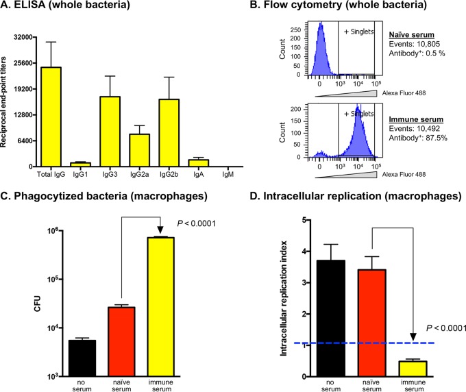
FIG 7.
ELISA, flow cytometry, and opsonophagocytic killing assays using WT B. mallei and immune serum (from mice vaccinated with the batA KO strain). (A) Immune serum samples were serially diluted and placed in duplicate wells of plates coated with whole paraformaldehyde-fixed B. mallei ATCC 23344 bacteria. Alkaline-phosphatase-conjugated goat anti-mouse isotype-specific antibodies were used for detection. The results are expressed as mean (plus standard error) reciprocal endpoint titers of 5 independently generated batches of immune serum. Naive serum was used to establish background reactivity. (B) Whole paraformaldehyde-fixed B. mallei ATCC 23344 bacteria were incubated with immune serum, labeled with a goat anti-mouse antibody conjugated to Alexa Fluor 488, and analyzed using a BD LSRII flow cytometer. The number of cells analyzed and the percentage binding antibodies on their surfaces are shown. Bacteria incubated with naive serum were used as controls. (C and D) Freshly grown B. mallei ATCC 23344 bacteria were incubated with immune serum for 30 min at 37°C, and the opsonized organisms were used to infect 2 wells of duplicate tissue culture plates seeded with murine J774 macrophages. (C) The infected cells were incubated for 1 h at 37°C to allow phagocytosis of the bacteria, washed, and treated with antibiotic for 2 h. Cells from one tissue culture plate were lysed, diluted, and plated onto agar medium to determine the number of bacteria phagocytized. The results are expressed as the mean numbers of CFU plus standard errors. (D) The other tissue culture plate was incubated for an additional 7 h, after which the cells were washed, lysed, diluted, and spread onto agar plates to calculate the number of intracellular organisms. The results are expressed as the mean (plus standard error) intracellular replication index, which was calculated by dividing the number of intracellular bacteria at the endpoint of the assay (second tissue culture plate) by the number of bacteria phagocytized (first tissue culture plate); an index below 1 (blue dashed line) indicates intracellular killing of bacteria. The assays were performed on 15 separate occasions. Bacteria incubated with PBS and naive serum (prior to infecting macrophages) were used as controls. The P values indicate that the differences observed between bacteria incubated with naive and immune sera were statistically significant using a paired t test.

