FIG 3.
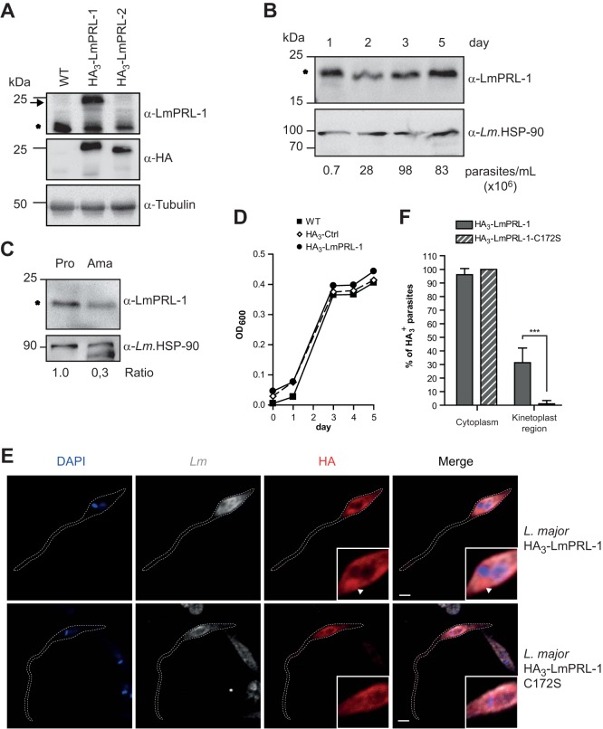
Expression and subcellular localization of LmPRL-1 in L. major parasites. (A) Detection of LmPRL-1 protein in promastigotes. Protein extracts were prepared from stationary-phase promastigote cultures of WT L. major and parasites ectopically expressing HA3-LmPRL-1 or HA3-LmPRL-2. The proteins were analyzed by immunoblotting with rabbit serum anti-LmPRL-1, as well as anti-HA antibody to estimate the specificity of the rabbit serum and with anti-tubulin antibody (loading control). Endogenous LmPRL-1 (expressed by WT parasites) is indicated by the asterisk and HA3-tagged proteins of transfected L. major by the arrow. (B) Expression of LmPRL-1 in logarithmic- or stationary-growth-phase promastigotes in vitro. A culture of WT L. major promastigotes in modified complete Schneider's medium inoculated with 1.0 × 10⁵ parasites/ml was analyzed daily for its concentration (parasites per milliliter) until it reached stationary phase (day 5). Expression of endogenous LmPRL-1 (marked by the asterisk) was analyzed by anti-LmPRL-1 immunoblotting, using the HSP-90 housekeeping gene as a control. (C) Expression of LmPRL-1 in promastigotes and amastigotes. Protein extracts prepared from the same number (20 × 106) of in vitro-cultivated promastigotes (Pro) and ex vivo-isolated amastigotes (Ama) were loaded onto SDS-PAGE. Expression of endogenous LmPRL-1 (marked by the asterisk) and of the HSP-90 housekeeping gene was analyzed by immunoblotting. The ratios of the band intensities are indicated below. (D) Ectopic expression of HA3-LmPRL-1 has no effect on in vitro growth of L. major promastigotes. WT promastigotes and parasites either carrying the empty pCLN-3xHA vector or expressing HA3-LmPRL-1 were cultivated in modified complete Schneider's medium starting at 1.0 × 10⁵ parasite/ml until stationary phase. Growth was monitored by measuring the OD600. (E and F) Subcellular localization of LmPRL-1 in L. major promastigotes depends on its C-terminal farnesylation. (E) Parasites expressing HA3-LmPRL-1 or HA3-LmPRL-1-C172S were analyzed by confocal microscopy after staining with an anti-HA antibody (red), an anti-Leishmania serum (gray), and DAPI (blue). The arrowheads indicate the region of the kinetoplast. Bars, 2 μm. (F) Means and standard deviations of more than 250 promastigotes per strain of L. major. ***, P < 0.001. The data presented are from one of three experiments with similar results.

