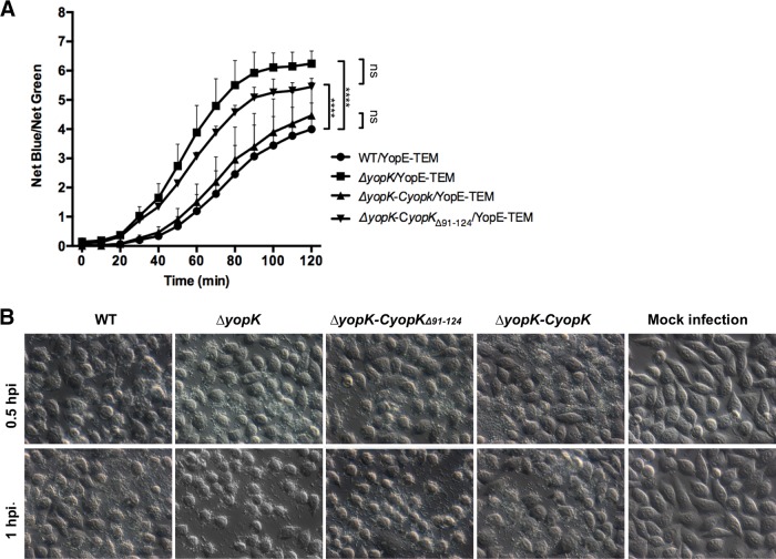
FIG 5.
The ΔyopK strain expressing YopKΔ91–124 exhibits Yop hypertranslocation. (A) The wild-type, ΔyopK, ΔyopK-CyopK, and ΔyopK-CyopKΔ91–124 strains harboring plasmid pBBR1-yopE-TEM were used in this experiment, in which the expression of YopE fused with a C-terminal TEM is driven by the intrinsic yopE promoter. The strains were cultured at 26°C in BHI medium and shifted to 37°C for an additional 3 h of incubation for the full induction of the T3SS. HeLa cells at 70 to 80% confluence were infected with the aforementioned strains at an MOI of 20. After 1 h of infection, cells were loaded with 1 μg ml−1 CCF2-AM substrate solution, and the plates were incubated at 37°C and analyzed with SpectraMax M5. The means and standard deviations (SD) are indicated. ****, P < 0.0001; ns, not significant (as determined by two-way ANOVA with Tukey's test for multiple pairwise comparisons). (B) HeLa cells were infected with different Y. pestis strains or mock infected as indicated. After 1 h of infection, DIC images of the infected cells were photographed under a Zeiss Axiovert 40 CFL microscope (Carl Zeiss, Germany).

