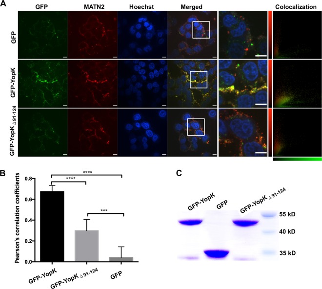
FIG 7.
YopK colocalizes with the endogenous MATN2 on the HeLa cell surface. (A) Equal amounts of purified GFP-YopK, GFP-YopKΔ91–124, or GFP were added into HeLa cells and incubated at 4°C overnight. Endogenous MATN2 on the HeLa cell surface was visualized using a rabbit anti-MATN2 antibody and a donkey anti-rabbit IgG secondary antibody conjugated to Alexa Fluor 555. All scale bars represent 9 μm. Images were acquired using an UltraVIEW Vox live-cell imaging system, and Pearson's correlation coefficient was used to quantify the degree of colocalization between the two channels (red and green) in a confocal image by using Volocity 6.1 software. (B) The bar graphs were drawn to show average Pearson's correlation coefficients (with error bars) from multiple images (n = 7 for each treatment) of the HeLa cells incubated with different proteins. One-way ANOVA with Bonferroni's multiple-comparison test was performed to analyze the significance of difference in bacterial adhesion between the different treatments (***, P < 0.001; ****, P < 0.0001). (C) SDS-PAGE analysis of the purified proteins confirmed that equal amounts of GFP-tagged YopK, YopKΔ91–124, or GST protein were added to cell cultures.

