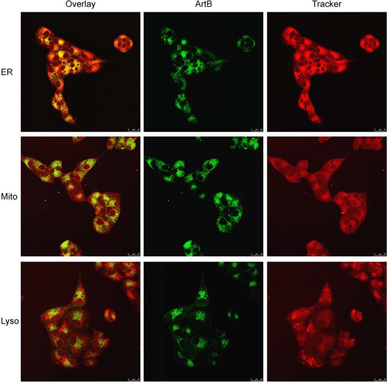
FIG 9.
Colocalization of ArtB with ER, mitochondrial, and lysosomal markers at 16 h. Vero cells were incubated with DyLight 488-labeled ArtB (10 μg/ml) for 16 h, with 2.5 μM ER-Tracker Red, 400 nM MitoTracker Red, or 250 nM LysoTracker Red being added for the final 30 min, after which cells were fixed and then examined by confocal microscopy as described in Materials and Methods. Separate ArtB and Tracker channels are shown, as is an overlaid image (nuclei unstained) (bar, 25 μm).

