Figure 3.
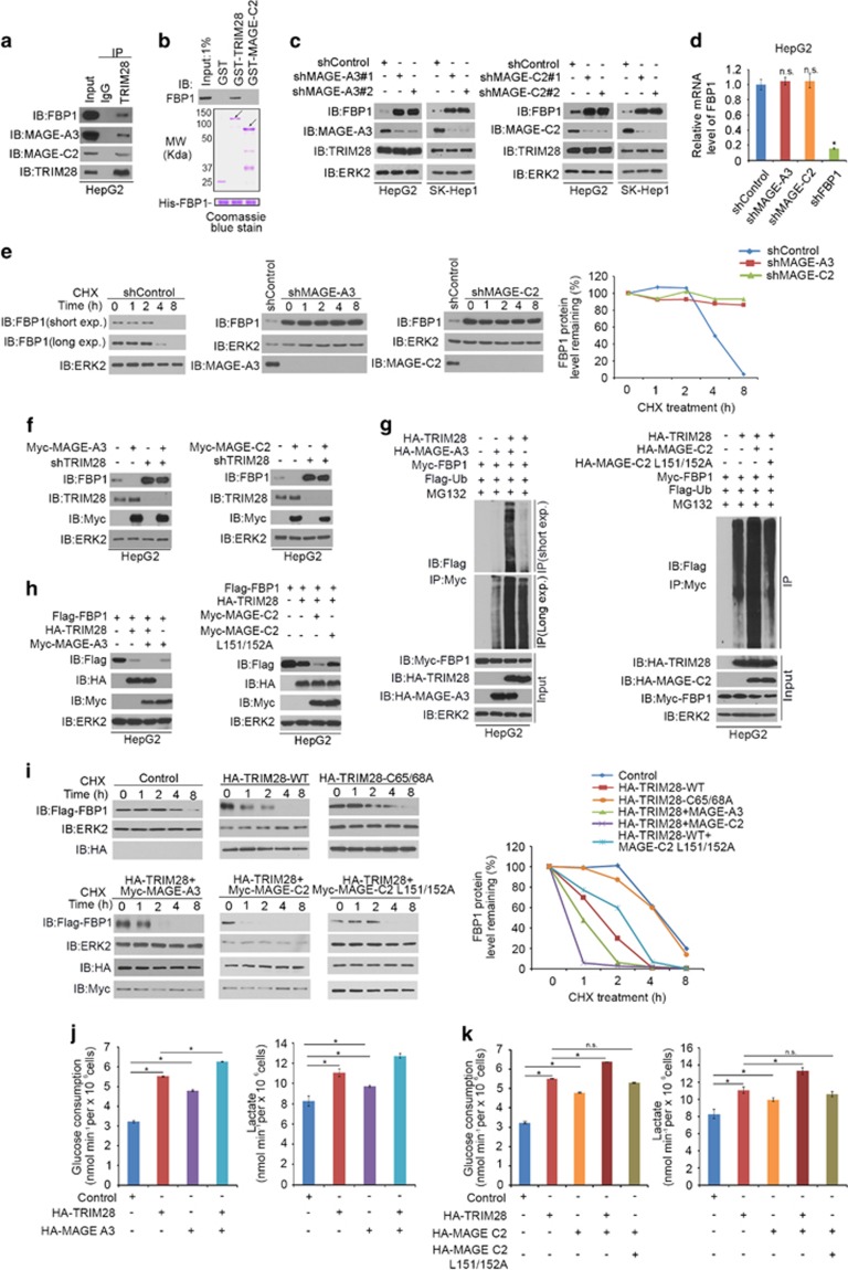
MAGE family proteins enhance FBP1 degradation by forming E3 ligase complexes with TRIM28. (a) HepG2 cell lysates were prepared for co-immunoprecipitation with anti-TRIM28 antibody followed by western blot analysis. (b) Bacterially expressed His-FBP1 proteins and GST, GST-TRIM28 or GST-MAGE-C2 recombinant proteins were subjected to in vitro protein binding assays followed by western blot analysis. Input samples were analyzed by Coomassie blue staining. (c, d) HepG2 and SK-Hep-1 cells were infected with control or two independent MAGE-A3 or MAGE-C2-specific shRNAs. After 48 h, cells were harvested for western blot (c) and reverse transcriptase (RT)–quantitative PCR (qPCR) analysis (d). (e) HepG2 cells were infected with control and MAGE-A3 and MAGE-C2 specific shRNAs. After 48 h, cells were treated with 50 μg/ml cycloheximide (CHX). At different time points, cells were harvested for western blot analysis. Exp., exposure. (f) HepG2 cells were infected with control and TRIM28-specific shRNAs. After 24 h, HepG2 cells were transfected with indicated constructs for another 24 h followed by western blot analysis. (g) HepG2 cells were transfected with indicated constructs for 24 h followed by western blot analysis. (h) HepG2 cells were transfected with indicated constructs for 20 h followed by treatment with 20 μM of MG132 for 8 h. Immunoprecipitated Myc-FBP1 proteins were analyzed by western blot. (i) HepG2 cells were transfected with control or indicated plasmids. After 24 h, cells were treated with 50 μg/μl CHX. At indicated time points, cells were harvested for western blot analysis. (j, k) Glucose consumption and l-lactate production were measured in the spent medium of HepG2 cells 48 h after transfected with indicated constructs. *P<0.01; NS, not significant.

