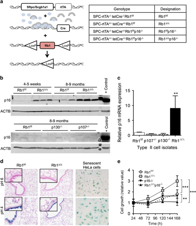
Figure 1.
p16 suppression is a unique RB1 pocket protein function in the lung epithelium in vivo with p16 induction after RB1 loss functioning to enhance lung epithelial cell growth. (a) Sftpc/Scgb1a1-rtTA+/−; tetCre+/− double transgenic mice were bred to Rb1f/f and p16−/− mice. Doxycycline (blue ovals) activates the rtTA (gray arches) leading to Cre-mediated Rb1 ablation in the lung epithelium. Genotypes resulting in the designated Rb1 and p16 status are indicated in the table. (b) p16 protein was induced in Rb1Δ/Δ lungs from mice at 4–5 weeks of age with increased expression sustained in lungs from 8 to 9-month-old mice by western blot analysis. p16 protein was not induced in p107−/− or p130−/− lungs (n=4–9 mice per group). Lysates were evenly loaded as assessed by reprobing for β-Actin (ACTB). A 3T3 cellular lysate served as a positive p16 control. (c) p16 messenger RNA was induced in Rb1Δ/Δ, but not p107−/− or p130−/−, type II cells as compared to Rb1f/f controls by quantitative reverse transcription PCR (mean±s.d.; n=3–4 isolates per group; **P<0.01). (d) p16 induction in Rb1Δ/Δ lungs was not associated with cellular senescence as assessed by senescence-associated β-galactosidase (SA-β-gal) activity at pH 6. Positive controls include SA-β-gal activity in senescent HeLa cells and lysosomal β-gal staining at pH 4 (n=3–4 mice per group at 9 months of age). Original magnifications: × 20 and insets × 40. (e) Growth of Rb1Δ/Δ primary type II cells in culture was increased as compared to Rb1f/f controls with additional loss of p16 reducing Rb1Δ/Δ type II cell growth to below Rb1f/f control levels. p16−/− type II cell growth was similar to Rb1/p16-proficient Rb1f/f controls (mean±s.d.; n=5–7 wells from 2 to 3 independent cell isolates; **P<0.01; ***P<0.001).

