Figure 3.
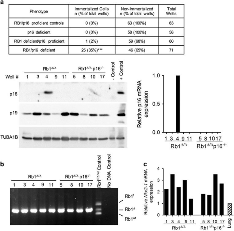
p16 suppresses immortalization of RB1-deficient cells. (a) Summary of percentage of tissue culture plate wells seeded with Rb1f/f, Rb1Δ/Δ, Rb1f/fp16−/− and Rb1Δ/Δp16−/− primary type II cells that resulted in immortalized cell populations (data are representative of three independent cell isolates and experiments; ***P<0.001 by Fisher’s exact test for RB1/p16 deficient vs all other groups). Representative western blot (left) and quantitative reverse transcription PCR (RT-PCR; right) analyses demonstrate that only 1 out of 5 immortalized cell populations derived from Rb1Δ/Δ type II cell cultures retained p16 expression and are thus included as RB1/p16 deficient in the summary. p19 was expressed in all cell populations. Lysates were evenly loaded as assessed by reprobing for TUBA1B. p16−/− lung and p16-positive mouse tumor tissue served as negative and positive controls, respectively. Well # represents distinct cell populations derived from individual wells. (b) Representative PCR analysis on DNA from Rb1Δ/Δp16−/− immortalized cells demonstrating all immortalized cells populations had Rb1 recombination (Rb1Δ) without detection of floxed (Rb1f) or wild-type (Rb1wt) alleles confirming derivation from Rb1 ablated lung epithelium. (c) All immortalized type II cell populations expressed the lung epithelial cell marker, Nkx2-1, at similar or higher levels than lung tissue as assessed by quantitative real-time RT-PCR.

