Figure 4.
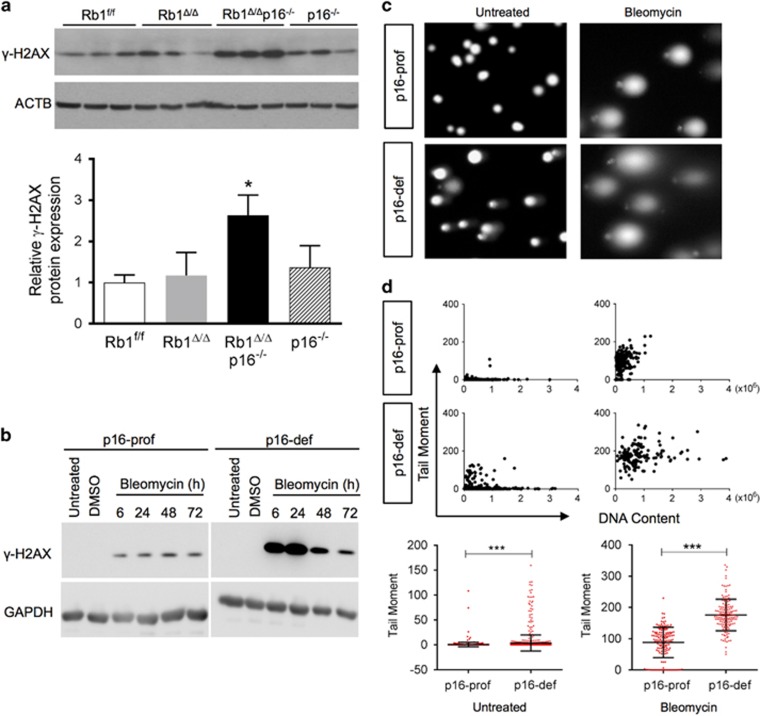
p16 protects RB1-deficient cells from DNA damage. (a) Expression of the DNA damage marker, γ-H2AX, was increased in Rb1Δ/Δp16−/− type II cell isolates as compared to Rb1f/f, Rb1Δ/Δ and p16−/− cells by western blot analysis with quantification by relative densitometric values normalized to ACTB. Lysates were evenly loaded as assessed by reprobing for ACTB (mean±s.d.; n=3 isolates from two independent experiments per group; *P<0.05). (b) γ-H2AX induction after bleomycin treatment was higher in p16 deficient (p16-def) as compared to p16-proficient (p16-pro) cells by western blot analysis of immortalized lung epithelial cells treated with bleomycin for 6, 24, 48 and 72 h. Untreated and DMSO vehicle-treated controls at the 6 h time point are shown. Cell lysates were evenly loaded as assessed by reprobing for GAPDH (representative of three independent experiments). (c) Representative comet assay SYBR Green stained images of p16-prof and p16-def cells untreated or treated with 10 μU bleomycin for 24 h. (d) Quantification of DNA damage by comet assay tail moments. Scatter plots (top) with quantification (bottom) of untreated and bleomycin-treated p16-prof and p16-def cells (mean±s.d.; n=920 total untreated cells and 140–150 representative treated cells per group from three independent experiments; ***P<0.001).

