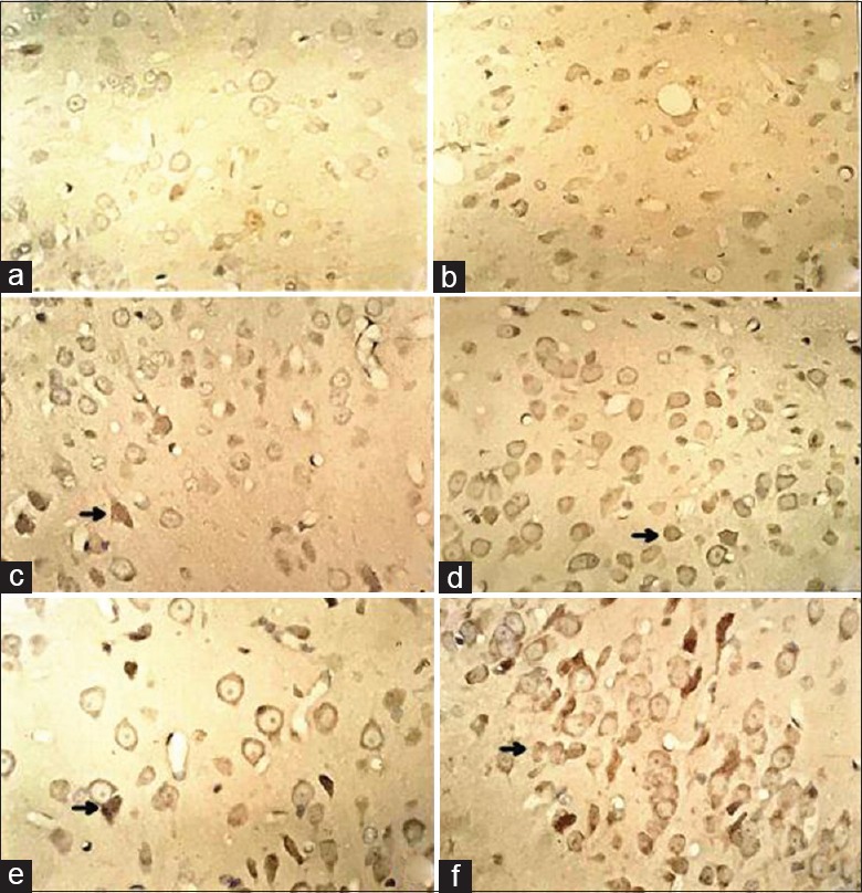
Figure 2.

Immunohistochemical staining results of HIF-1α expression at each time point in Ad and AdHIF-1α groups. HIF-1α-positive cells with brownish yellow granules as arrows indicate were HIF-1α expressions (original magnification, ×400). Rare brownish yellow granules were found in CIR group. (a) HIF-1α expression in CIR group at 6 h postreperfusion; (b) HIF-1α expression in AdHIF-1α group at 6 h postreperfusion; (c) HIF-1α expression in CIR group at 24 h postreperfusion; (d) HIF-1α expression in AdHIF-1α group at 24 h postreperfusion; (e) HIF-1α expression in CIR group at 72 h postreperfusion; (f) HIF-1α expression in AdHIF-1α group at 72 h postreperfusion. HIF-1α: Hypoxia-inducible factor-1α; Ad: Recombinant adenovirus; AdHIF-1α: Recombinant adenovirus vector containing HIF-1α; CIR: Cerebral ischemia and reperfusion.
