
Figure 1.
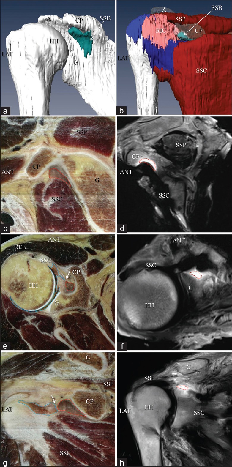
Three-dimensional shape and topographical location of the SSB: Sectional anatomy of SSB and its adjacent structures in the CVH sections and MR images. (a, b) The three-dimensional shape of SSB, the spatial relationship between SSB and its adjacent structures. The SSB are shown in the sagittal oblique CVH (c) and MR images (d); the axial CVH (e) and MR images (f); the coronal oblique CVH (g) and MR images (h). Arrow in e and g: Spectrum separating the SSB and GHAC (in the area marked by blue dashed line) is shown. Area marked by red dashed line: SSB; A: Acromion; ANT: Anterior side; C; Clavicle; CP: Coracoid process; G: Glenoid; GHAC: Anterior part of the glenohumeral articular cavity; HH: Humeral head; LAT: Lateral side; RIC: Rotator interval capsule; SSB: Subscapular bursa; SSC: Subscapularis; SSP: Supraspinatus; SUP: Superior side; MR: Magnetic resonance; CVH: Chinese visible human.