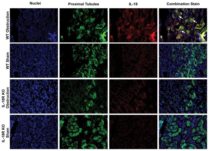
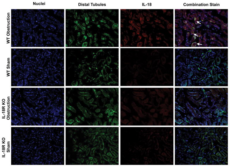
Figure 3. Dual labelled immunofluorescent staining for IL-18 and tubular epithelial cells following UUO.
A. Photographs (magnification 400X) depicting renal cortical IL-18 production (red) and proximal tubular staining (Lotus Tetragonolobus Lectin; green) in WT and IL-18R KO animals exposed to sham operation or one week of UUO. B. Photographs (magnification 400X) depicting renal cortical IL-18 production (red) and distal tubular staining (Peanut Agglutinin; green) in WT and IL-18R KO animals exposed to sham operation or one week of UUO. White arrows indicate IL-18 staining overlying tubules.


