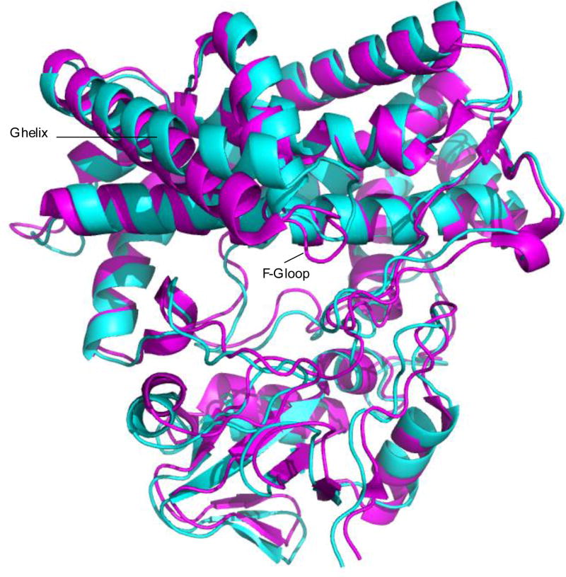
Figure 5.
Backbone superposition of 2Y98 [purple) and REP1 (cyan) structures of M-IV-bound MycG. The RMS deviation of the backbone atom positions of the structures is 1.49 Å. The largest differences are seen in the F-G loop and G helix (marked) with a concerted displacement of the β-rich region (towards the bottom of the structures as shown). Structures are shown in the same orientation as in Figure 2.

