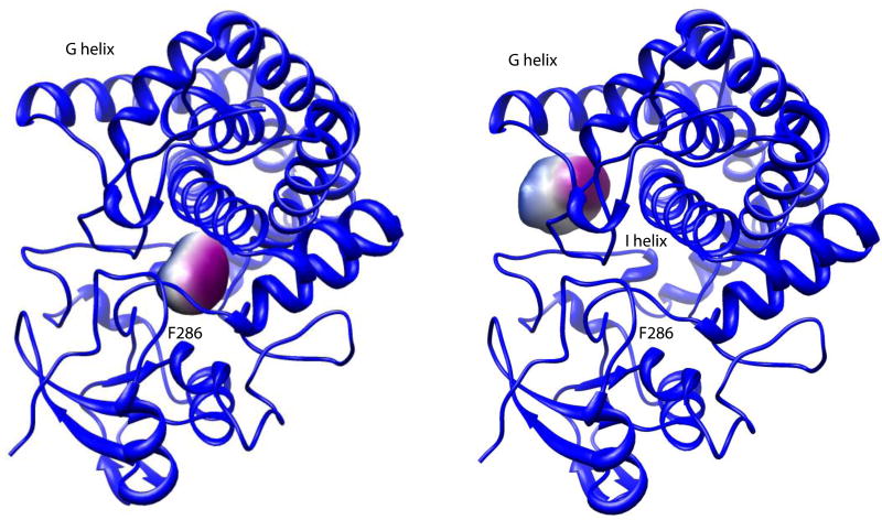
Figure 6.
Active site voids upon vacating M-IV from 2Y98. Two voids are detected, with the larger void (right) adjacent to the G helix and the F-G loop. The smaller void (left) is between the I helix and primary substrate contact Phe 286, immediately preceding the start of the β3 strand. Both figures are from the same perspective. Figures generated by V3 program (3vee.molmovdb.org). Void coloring is for visualization only. See text for details.

