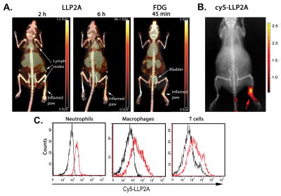
FIGURE 1. 64Cu-LLP2A and 18F-FDG are retained in inflamed mouse paws following CFA injection.
(A) PET/CT comparison of 64Cu-LLP2A and 18F-FDG uptake in injected (right) and uninjected (left) mouse paws. PET signal is indicated in Table 1. Note that 64Cu-LLP2A is taken up by lymph nodes distal to the site of injection whereas lymph nodes are not visible in the 18F-FDG-probed animal at 45 min post injection. The difference in scan times for 64Cu-LLP2A and 18F-FDG scan times reflects the different pharmacokinetics of these probes. (B) In vivo fluorescence image showing accumulation of LLP2A-Cy5 in the injected paw (right) compared to uninjected paw (left). (C) Flow cytometry on cells collected from the inflamed paw from animal injected with LLP2A-Cy5 or not (control) demonstrates neutrophils, macrophages, and T cells stain positively with Cy5-labeled LLP2A (red lines). Black lines represent control animals not injected with LLP2A-Cy5.

