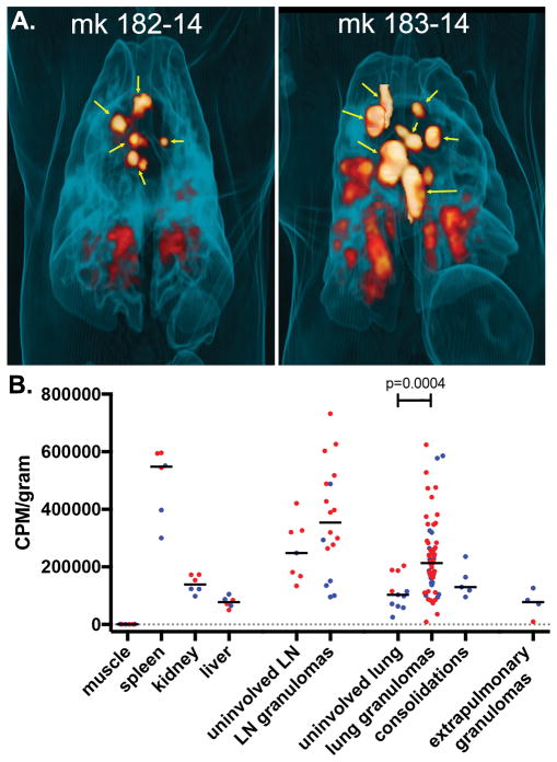
Figure 4. Uptake of 64Cu-LLP2A by cynomolgus macaque tissues.
(A) 3-D rendered maximum intensity projections showing overall 64Cu-LLP2A uptake in granulomas and infected lymph nodes (non-thoracic uptake removed) before necropsy. Yellow arrows indicate lymph nodes. (B) Quantitative analysis of LLP2A uptake by tissues obtained at necropsy. CPM/gram data are normalized to tissue mass and markers indicate tissues from monkeys 182-14 (blue) and 183-14 (red).

