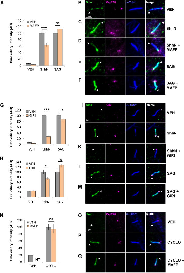
Figure 2.
cPLA2 influences ShhN-, but not SAG-induced Smo ciliary translocation. For all panels *** indicates p ≤ 0.0001; * indicates p ≤ 0.05; ns indicates p > 0.05; nt, not tested. Error bars indicate SEM. A–M. NIH3T3 cells pretreated with MAFP (5 µM) or GIRI (5 µM) were stimulated with ShhN conditioned media or SAG (100 nM) and imaged by immunofluorescence confocal microscopy. Ciliary tips are indicated by arrowheads. Smo and/or Gli3 signal in primary cilia was quantified by counting ≥100 cells over a minimum of 2 experiments. Significance indicated in A, G and H was calculated based upon total cell number analyzed using a two-way ANOVA. N–Q. Smo localization was quantified as above in NIH3T3 cells treated with cyclopamine (10 µM) +/− MAFP (5 µM). Significance was determined using a two-tailed Student’s t-test. For all cilia shown, Smo is green, Gli3 is magenta, the ciliary marker acetylated α-tubulin is blue and the ciliary base marker Cep290-GFP is magenta.

