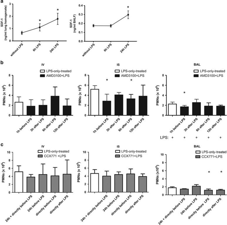
Figure 1.
Effect of our model on SDF-1 levels in the lungs of mice (a). Mice inhaled LPS and SDF-1 levels were determined in the lungs (n=8) and BAL (n=4). Data are presented as mean ±S.D.; *P<0.05 versus without LPS. Time optimum for the administration of the CXCR4- (b) and CXCR7-antagonist (c). The inhibitors were given at indicated time points and, 24 h after LPS-inhalation, migration of PMNs into the different compartments of the lung (IV=intravascular; IS=interstitial; BAL=bronchoalveolar lavage) was evaluated. Data are presented as mean ±S.D.; n≥4; *P<0.05 versus LPS-only treated

