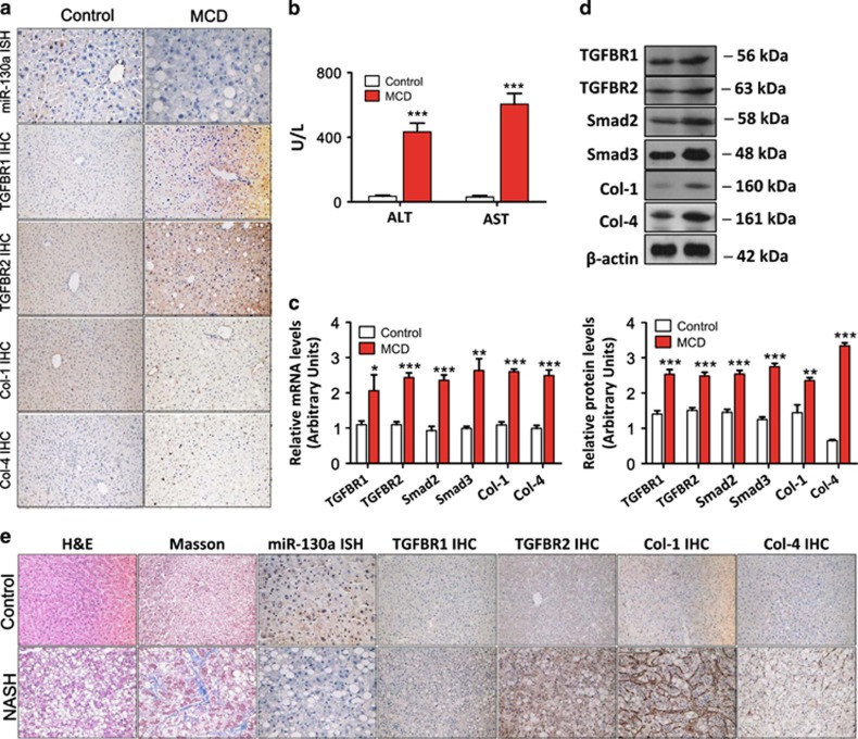
Figure 3.
Validation of the differential expression of miR-130a-3p in the livers of mice and patients. (a) miR-130a-3p expression was decreased, and the expression of TGFBR1, TGFBR2, Col-1, and Col-4 was increased in the liver tissues of the MCD-fed mice. TGFBR1, TGFBR2, Col-1, and Col-4 expression was detected by IHC ( × 200 magnification), and miR-130a-3p expression was detected using an ISH ( × 400 magnification) assay. Positive staining is indicated by a brown color. (b) Effects of the MCD diet on serum ALT and AST levels. Values represent the mean±S.D., ***P<0.001 compared with control. (c) Hepatic mRNA and (d) protein expression of TGFBR1, TGFBR2, Smad2, Smad3, Col-1, and Col-4 were upregulated in the MCD-fed mice compared with the control mice. β-actin was used as a loading control. Values represent the mean±S.D., *P<0.05, **P<0.01, ***P<0.001 compared with control. (e) Histopathological changes were evaluated with hematoxylin and eosin staining, and Masson’s trichrome staining. miR-130a-3p expression was decreased, and TGFBR1, TGFBR2, Col-1, and Col-4 levels were increased in the liver tissues of patients with NASH-related liver fibrosis. The expression of miR-130a-3p was detected by ISH ( × 400 magnification), and TGFBR1, TGFBR2, Col-1, and Col-4 were detected using an IHC ( × 200 magnification) assay. Positive staining is indicated by a brown color

