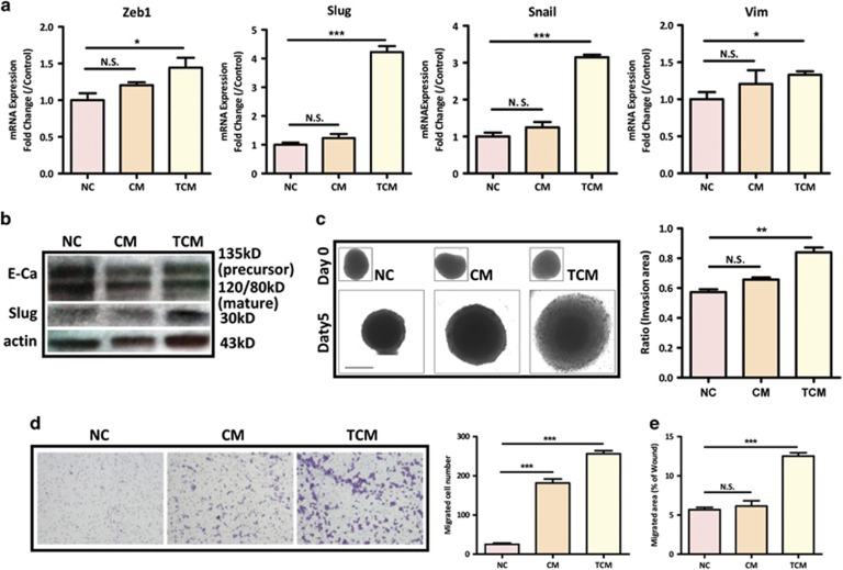
Figure 2.
hMSCs promote metastatic phenotype of colon cancer cells. (a) After incubation with CM or TCM, the expression levels of EMT-related genes in HT29 were evaluated by quantitative PCR. Data are presented as the means±S.D. n=3. NS, no significance. *P<0.05, ***P<0.001 versus control; (b) Western blot analysis showed that CM and TCM decreased the expression of E-cadherin, whereas TCM increased the expression of Slug; (c) Invasion ability of HT29 treated with CM or TCM was evaluated by 3D spheroid invasion assay (scale bar, 500 μm). Invasion ratio=((area D5−area D0))/(area D0). The experiment was repeated three times, **P<0.01 versus control group; (d) Cell migration was determined by transwell assay in SW1116. 1 × 10⁴ SW1116 cells were seeded in the upper chamber whereas CM or TCM were administrated in the lower chamber. The experiment was repeated three times. ***P<0.001 versus control group; (e) wound healing assay was used to determine cell migration. Quantification data was presented as mean±S.D. from three independent experiments, ***P<0.001 versus control group

