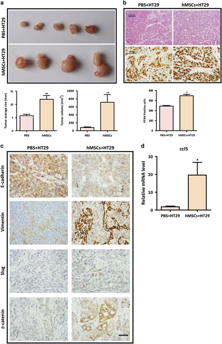
Figure 6.
hMSCs promote CRC development and EMT in vivo BALB/C mice were subcutaneously injected with 1 × 10⁷ HT29 cells mixed with 1x10⁷ hMSCs (n=4) or alone (n=5). (a) The tumors were excised after 2 weeks, and tumor average size and tumor volume in xenograft model were measured. **P<0.01, versus PBS group; (b) H&E staining and immunohistochemical staining for PCNA (proliferating cell nuclear antigen) of xenograft tumors from either HT29 alone or co-injection group; quantification data are from at least five high fields. *P<0.05. Scale bar, 50 μm and 100 μm; (c) immunohistochemical staining for EMT markers (E-cadherin and Vimentin), β-catenin and Slug. Scale bar, 100 μm; (d). The expression of CCL5 is increased in nude mice injected with HT29 and hMSCs. Tumor tissues were collected from mice injected with HT29 alone or HT29 with hMSCs. RNA was extracted and determined for expression of CCL5 by real-time PCR. Data are presented as the means±S.D. n=3 *P<0.05 versus control

