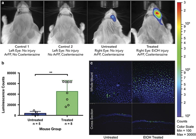
Figure 6.
(a) IVIS images comparing mice from groups control 1 (addition of protein with no injury or substrate), control 2 (addition of substrate with no injury or protein), untreated (addition of protein and substrate with no injury), and treated (addition of protein and substrate following corneal injury). (b) Comparison of bioluminescence data from the total cohort (n=13) of mice receiving ethanol treatment (treated, n=8) or PBS (untreated, n=5). (c) TUNEL analysis of corneal sections. Whole mount (top) and cross-section (bottom) images of corneal tissues from untreated (PBS, left) and treated (ethanol, right) mice indicating the presence of apoptotic (green) and non-apoptotic (blue) nuclei

