Figure 3.
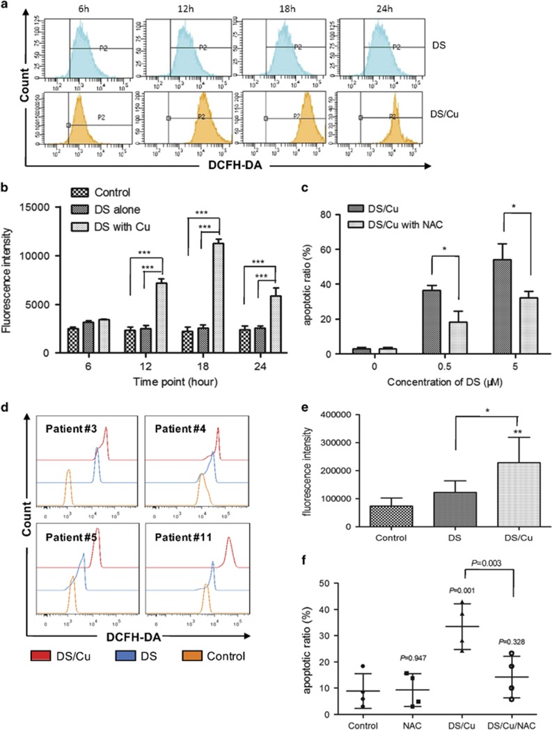
DS/Cu-induced cytotoxicity is associated with intracellular ROS accumulation in leukemia stem-like cells. (a) CD34+/CD38− KG1a cells were treated with DS (0.5 μM) ± Cu (1 μM) for the indicated intervals. Intracellular ROS level was determined by analysis of DCFH-DA fluorescence intensity. (b) Histogram of ROS generation in CD34+/CD38− KG1a. ***P<0.001. (c) Histogram of apoptosis percentage in CD34+/CD38− KG1a cells in the presence of free radical scavenger NAC (10 mM) ± DS/Cu for 24 h. *P<0.05. (d) Intracellular ROS level was examined by analysis of DCFH-DA fluorescence intensity. Primary CD34+ samples isolated from AML patient #3, #4, #5, #11 were treated with DS (0.1 μM) +/- Cu (1 μM) for 6 h. *P<0.05, **P<0.01. (e) Histogram of ROS generation in CD34+ primary cells (n=4). (f) Histogram of apoptosis percentage in CD34+ primary cells (n=4) after treated with DS/Cu or DS/Cu/NAC

