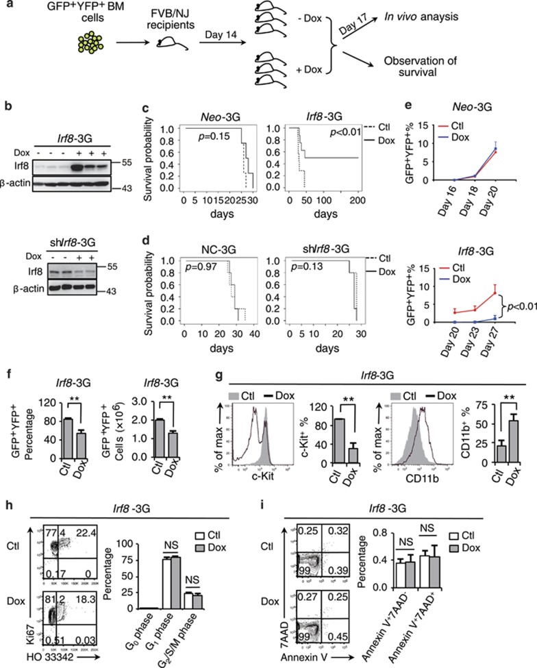
Figure 5.
Irf8 represents a potential oncorepressor in APL. (a) The experimental strategy used for the in vivo analyses of the APL mouse model genetically incorporated with a Tet-On3G gene-inducible expression. (b) Western blotting assay on the Irf8 protein level of APL cells (Irf8-3G, upper panel, or shIrf8-3G, bottom panel) with or without exposure to Dox administration (200 μg/ml in the drinking water) for 3 days. (c–d) Kaplan–Meier survival curves of the Irf8-3G mice (c) and shIrf8-3G mice (d) after treatment with or without Dox (n≥5), Neo-3G (n=4) and NC-3G (n=5) mice were used as the controls. (e) Dynamic monitoring of GFP+YFP+ APL cell percentages in the peripheral blood of the Neo-3G or Irf8-3G mice after exposure to Dox. (f–i) The Irf8-3G mice were treated with or without Dox for 3 days. (f) The percentages (left panel) and absolute numbers (right panel) of GFP+YFP+ APL cells in the total BM cells. (g) The flow cytometry analyses of the expressions of c-Kit and CD11b within the leukemic BM compartment. (h–i) Flow cytometry analyses of the cell cycle (h) and survival (i) of BM APL cells. All data in this figure are presented as the mean±S.D., *P<0.05, **P<0.01

