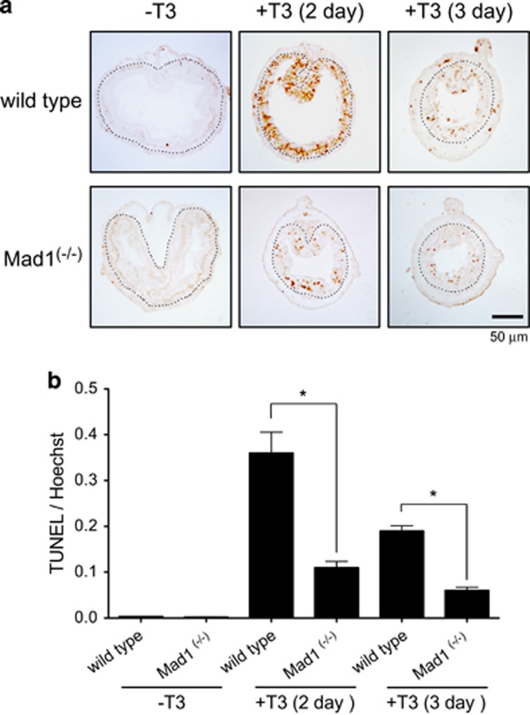
Figure 8.
Mad1 knockout inhibits cell death in the epithelium during T3-induced metamorphosis. (a) Cross-sections of the intestine of premetamorphic stage 54 tadpoles treated with 5 nM T3 for 0, 2 or 3 days were stained for apoptosis by TUNEL. Note that apoptosis in the epithelium peaked after 2 days of T3 treatment and that there were more intestinal epithelial cells in wild-type animals strongly stained by TUNEL compared with the ones in Mad1 (−/−) tadpoles. The dotted lines depict the epithelium–mesenchyme boundary. Scale bar, 50 μm. (b) Quantitative analysis of apoptosis by counting TUNEL-positive areas in the epithelium and normalized by the total cellular area in epithelium determined by Hoechst staining

