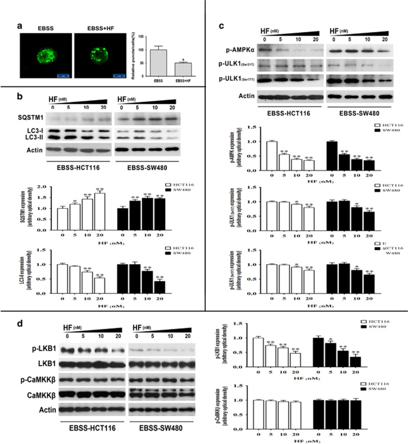
Figure 2.
HF inhibits autophagy in CRC cells under nutrient-poor condition. (a) Accumulation of GFP-LC3-II puncta in HCT116 cells with 20 nM HF for 2 h in EBSS medium. The distribution of GFP-LC3-II was examined by confocal microscope (left panel) and quantitative analysis (right panel). Scale bar: 10 μm. *P<0.05, **P<0.01. (b) Protein expressions of SQSTM1 and LC3-II in HCT116 and SW480 cells (upper panel); quantitative analysis of protein expressions (bottom panel) treated with 0, 5, 10, 20 nM HF for 2 h in EBSS medium. *P<0.05, **P<0.01. (c) Protein expressions of phospho-AMPKα, phospho-ULK1 at Ser317 and Ser777 in HCT116 and SW480 cells (upper panel); quantitative analysis of protein expressions (bottom panel) treated with 0, 5, 10, 20 nM HF for 2 h in EBSS medium. *P<0.05, **P<0.01. (d) Protein expressions of phospho-LKB1, phospho-CaMKKβ in HCT116 and SW480 cells (left panel); quantitative analysis of protein expressions (right panel) treated with 0, 5, 10, 20 nM HF for 2 h in EBSS medium. *P<0.05, **P<0.01

