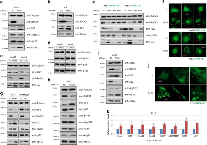
Figure 4.
RACK1 depletion-induced autophagy is not a cell-line-specific phenomenon. (a–d and g–i) Control or RACK1 siRNAs were transfected (20 pmol) into HeLa (a), HDF (b), HepG2 and Hep3B (c, d), MCF7 and MDA-MB231 (g), MEF (h) and U2OS (i) cells. After 48 h, immunoblot analysis was performed using the indicated antibodies. (e, f and j) Indicated siRNAs (50 pmol) were transfected into EGFP-LC3 stably expressing HepG2, Hep3B and U2OS cells, followed by fluorescence microscopy (f and j) and immunoblot analyses using the indicated antibodies (e). (k) In each cell line, the intensities of LC3 proteins were normalized against that of tubulin protein, and the relative expression in RACK1 siRNA-treated cells compared with that in control cells was plotted. *P<0.05, **P<0.01 (Student’s t-test)

