Figure 2.
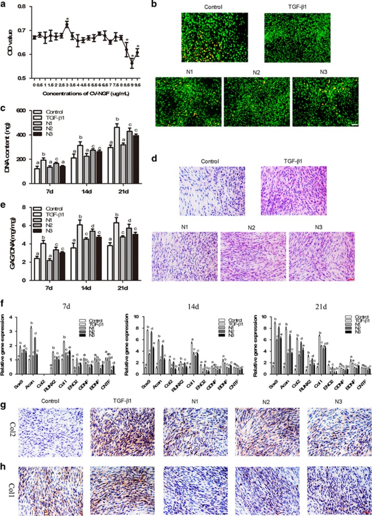
Effects of NGF on BMSCs in monolayer cultures. (a) The MTT assay was used to detect the cytotoxic effects of NGF on BMSCs. (The values are means±S.D., n=6, *P<0.05). (b) The effect of NGF on the viability of BMSCs cultured in monolayers for 21 days. Scale bar, 100 μm. (c) Quantification of the DNA content at 7, 14 and 21 days of culture. (d) HE staining was used to determine the morphology of BMCSs cultured in monolayers for 21 days. Scale bar, 100 μm. (e) GAG secretion from the cells after treatment for 7, 14 and 21 days. (f) qRT-PCR was used to determine the expression of the ACAN, SOX9, COL2A1, COL1A1, RUNX2, ENO2, GDNF, BDNF and CNTF genes on days 7, 14 and 21. (g and h) Immunohistochemistry. BMSCs were stained with COL1A1 (g) and COL2A1 (h) antibodies after 21 days in culture. Scale bar, 100 μm. The values are means±S.D., n=6; bars with different letters are significantly different from each other at P<0.05

