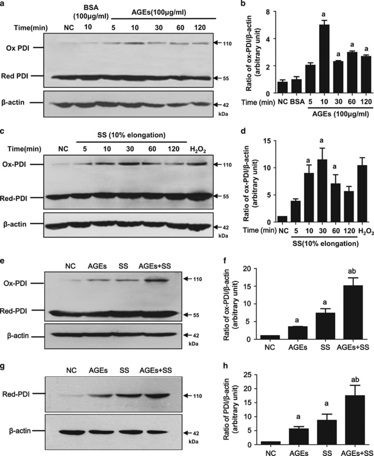
Figure 3.
SS and AGEs activate PDI in VSMCs in vitro. 70% confluent VSMCs were serum-starved for 24 h and treated with SS, AGEs or both for 10 min (SS, 10% elongation and indicated time) and (AGEs, 100 μg/ml)or for 1 h, and cultured for additional 23 h, and harvested for detection of ox-PDI or total PDI by western blot, respectively. (a, c and e) SS and/or AGEs-induced increases of ox-PDI in VSMCs; (g) SS and/or AGEs-induced increases of total PDI in VSMCs. Beta-actin was set as internal loading control; (b, d, f and h) statistical results of ratios of ox-PDI/ or total PDI/Beta-actin from (a, c, e and g) from three independent experiments. a, P<0.05 versus negative control (NC); b, P<0.05 versus AGEs or stretch stress (SS) by analysis of variance (ANOVA) with LSD test. Data are shown as the means±SEM

