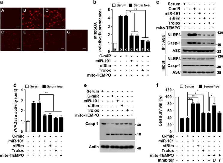
Figure 7.
MiR-101-3p blocks serum deprivation-induced mitochondrial ROS production, NLRP3 inflammasome activation, and caspase-1 activation. Cells were transfected with C-miR, miR-101 or siBim, followed by treatment with Trolox, Mito-TEMPO, Ac-DEVD-cho (D) or Ac-YVAD-cho (Y) in serum-free media for 12 h (ROS assay and IP), 24 h (caspase assay) or 30 h (MTT assay). (a) Mitochondrial ROS generation was identified by MitoSOX-based confocal microscopy. Scale bars, 50 μm. A: 5% FBS, B: serum-free, C: serum-free+100 nM C-miR, D: serum-free+100 nM miR-101, E: serum-free+100 nM siBim, f: serum-free+10 μM Trolox, g: serum-free+10 μM mito-TEMPO. (b) Fluorescence intensity was determined by Image J software. (c) NLRP3 inflammasome activation was determined by Western blotting following IP. YVADase activity (d) and proteolytic caspase-1 activation (e) were determined in cell lysates by colourimetric assay and Western blot analysis. (f) Cell viability was also determined by MTT assay. *P<0.05 and **P<0.01

