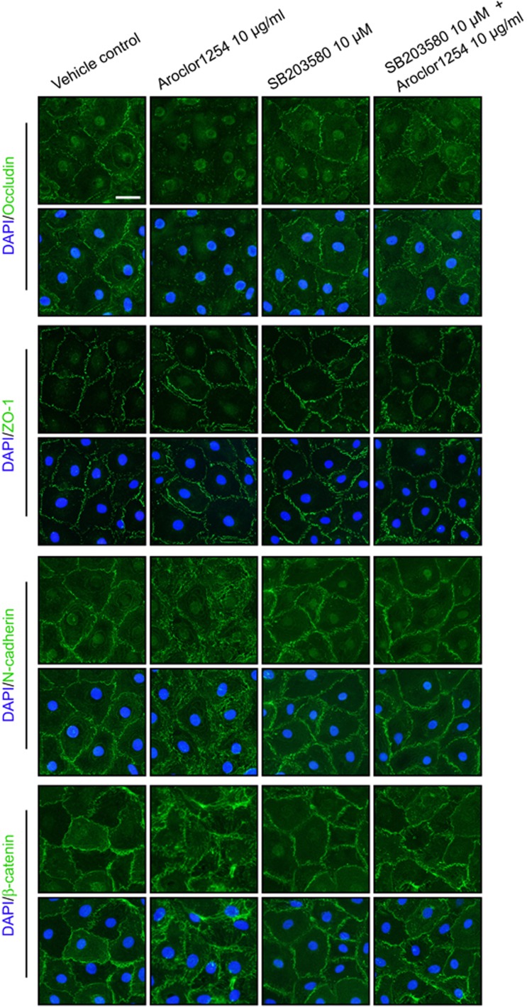
Figure 3.
Aroclor1254 influenced distribution of junction proteins in primary SCs. Primarily isolated SCs were cultured on matrigel-coated coverslips at 0.05 × 10⁶/cm2. On day 3, Aroclor1254 (10 μg/ml), SB203580 (10 μm), or the mixture of the above two was added to the medium. After 24 h, the cells were fixed with 4% paraformaldehyde. The distribution of BTB proteins at cell–cell junctions was observed by immunofluorescence using Alexa Fluor 488-conjugated secondary antibody (green) with cell nuclei stained with DAPI (blue). A reduced signal of occludin and disturbed distributions of N-cadherin and β-catenin were detected at cell junctions of Aroclor1254-treated SCs, as compared with the sharply defined signals in the control and the Aroclor1254+SB203580 groups. Scale bar=25 μm, which applied to all micrographs in Figure 3

