Figure 4.
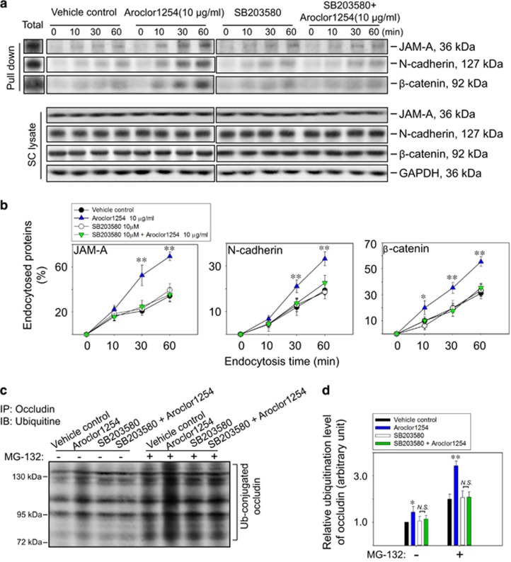
Aroclor1254 accelerated junction protein endocytosis and ubiquitination via p38 MAPK. (a) Immunoblot analysis of endocytosed JAM-A, N-cadherin, and β-catenin in SCs at different time points (0, 10, 30, 60 min) after cell surface biotinylation for 30 min in the presence of Aroclor1254 (or the mixture of Aroclor1254 and SB203580). The endocytosed proteins were pulled-down by UltraLink Immobilized NeutrAvidin Plus Resin after stripping residual biotin on cell membrane as described in ‘Materials and methods’ section. Total biotinylated proteins at 0 min without stripping were also detected as a positive control. Cell lysates without pull-down were also analyzed by immunoblot to confirm identical levels of the tested protein between groups with GAPDH served as the loading control. (b) Line and scatter graphs summarizing the result shown in a by calculating the percentage of endocytosed protein at each data point versus the total biotinylated protein. Each bar refers to mean±S.D. of n=3 independent experiments using SCs cultured from different rats. *P<0.05; **P<0.01, compared with the vehicle control at the same time point. (c) Co-immunoprecipitation assay using SCs lysates to detect the ubiquitination level of occludin after Aroclor1254 treatment or combined treatment of Aroclor1254 and SB20358 for 48 h. MG-132 of 50 μm was applied to inhibit the proteasome degradation of ubiquitine-conjugated proteins. (d) Histogram summarizing the result shown in c with the ubiquitinized occludin in the vehicle control SCs without MG-132 treatment was arbitrarily set at 1. *P<0.05; **P<0.01, compared with the corresponding control group. NS, no significant difference between two indicated groups

