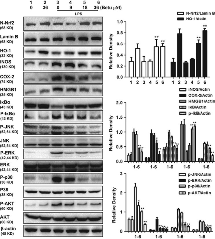
Figure 6.
Effect of betulin on anti-inflammatory and antioxidant signaling pathways in LPS-stimulated macrophages. RAW264.7 cells were plated in 6-well plates, preincubated with betulin (9, 18 and 36 μM) for 1 h, and then challenged with LPS (500 ng/ml) for 18 h. Cell lysates were immunoblotted with different antibodies. A representative Western blot is shown in the left panel. Shown in the right panel are means±S.E.M. of three independent experiments. *P≤0.05, **P≤0.01 versus the LPS group

