Figure 6.
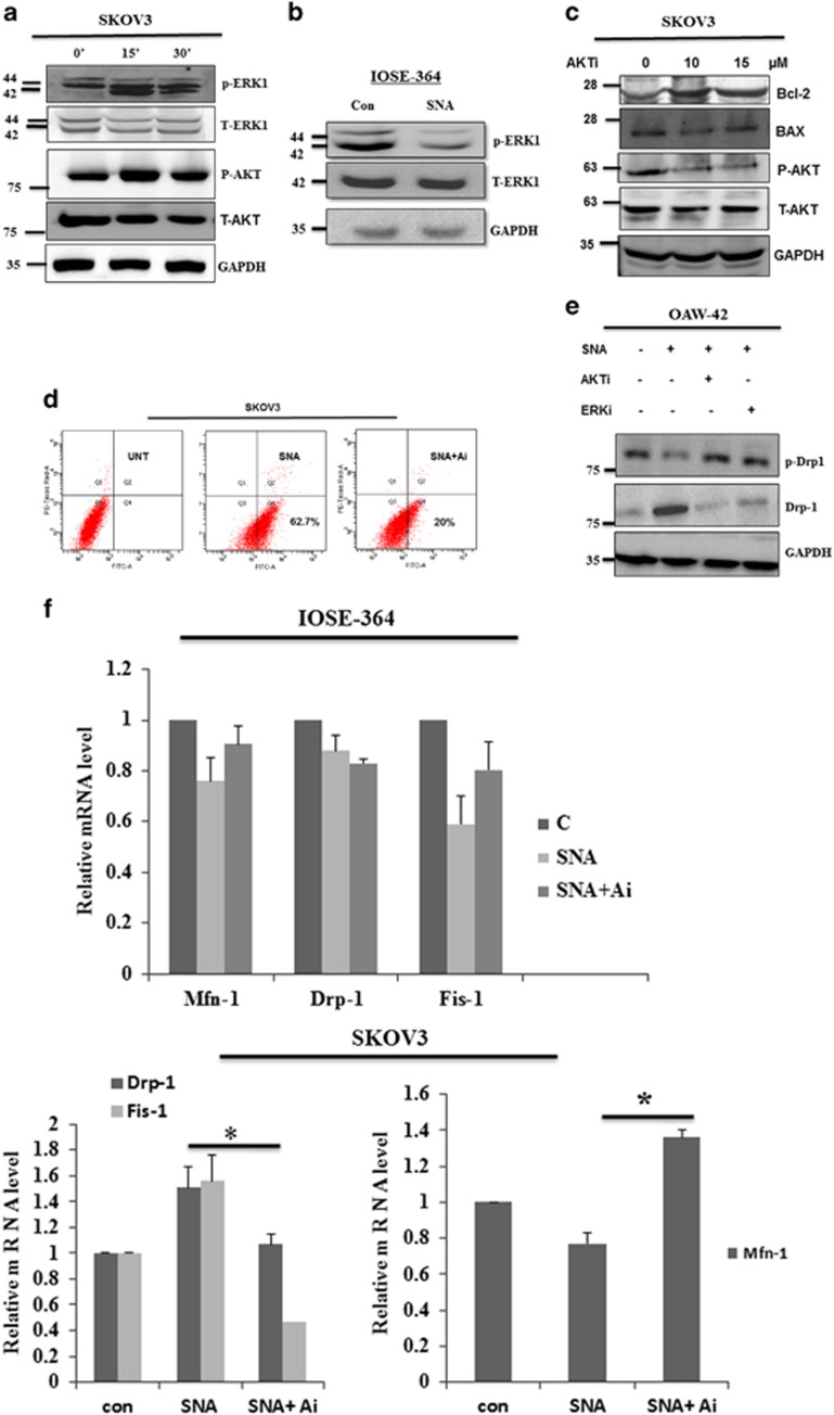
SNA induces apoptosis through activation of the AKT-ERK1/2 pathways. Cells were stimulated with 12 μg/ml of SNA for indicated time periods. (a and c) Lysates prepared from the SKOV3 cells were analyzed for p-AKT, p-ERK1, T-AKT, T-ERK1, Bax, Bcl-2 with GAPDH as loading control. (b) Lysates from IOSE-364 were checked for the expression of p-ERK1 after 30-min incubation and T-ERK1 after 24-h incubation with GAPDH as loading control. (d) Apoptotic induction in SKOV3 cells were quantified by flow cytometry after 24 h of SNA treatment in presence or absence of 10 μM AKTi by Flow cytometry. (e) OAW-42 cell lysates were analyzed for p-Drp-1 and Drp-1 after 30-min and 24- h incubation with SNA, respectively. GAPDH was used as loading control. (f) The Q-PCR of Mfn-1, Drp-1 and Fis-1 after 4 h of SNA treatment was observed in IOSE-364 and SKOV3 cells

