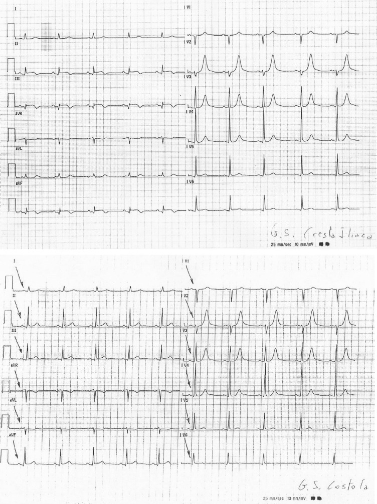
Figure 8.
Two different recorded electrocardiograms (EKGs) of the same patient at the same time, but with different positioning of the left leg lead. Note that only the positioning of the lead on the Iliac Crest can explore the inferior cardiac area. QRS voltage appears to be largely modified G.S. Cresta Iliaca, G.S. Iliac Crest; G.S. Costola, G.S. Rib.

