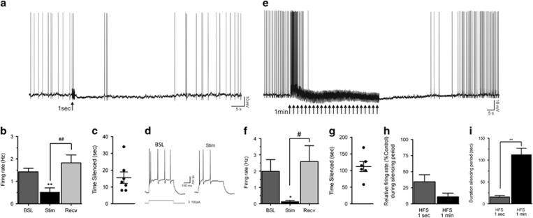
Figure 4.
Subthalamic nucleus (STN) activity following high-frequency stimulation (HFS). STN activity following a 1-s stimulation is represented in (a–d). (a) Continuous intracellular recording of a STN neuron in current-clamp mode. Tonic firing was depressed following a 1-s HFS (arrow), and the silencing period lasted 34 s until recovery to pre-HFS level. (b) On average, the action potential (AP) firing was significantly decreased from 1.43 to 0.52 Hz, with recovery to 1.52 Hz (n=7). **p<0.01 indicates significant effect of stimulation compared with baseline. ##p<0.01 shows significant effect of recovery compared with stimulation. Error bars indicate SEM. (c) Following delivery of a 1-s HFS, the silencing period lasted 16 s (n=7). (d) In another neuron, injection of a depolarizing current step before HFS (Control) triggered five APs, and the same current step delivered at the same holding potential during the silencing period (Stim) triggered only three APs. STN activity following a 1-min stimulation is represented in (e–i). (e) Continuous recording of a STN neuron. Delivery of a 1-min HFS (multiple arrows) first increased tonic firing for a few seconds and then markedly inhibited neuronal activity for 105 s until recovery to pre-HFS level. (f) On average, the AP firing was significantly decreased from 1.99 to 0.13 Hz, with recovery to 2.59 Hz (n=6). *p<0.05 indicates significant effect of stimulation compared with baseline. #p<0.05 shows significant effect of recovery compared with stimulation. Error bars indicate SEM. (g) Upon delivery of a 1-min HFS, the silencing period lasted 112 s (n=6). (h) The depressing effect of HFS on neuronal activity was more pronounced with a 1-min duration (89% decrease) compared with 1-s (66% decrease). (i) The duration of the silencing period was significantly longer with a 1-min HFS compared with a 1-s HFS. **p<0.01 indicates significant effect of 1-min stimulation compared with 1-s stimulation. Error bars indicate SEM. BSL, baseline; Recv, recovery; Stim, stimulations.

