
Fig. 4. Effect of nitration on microvesicle release.
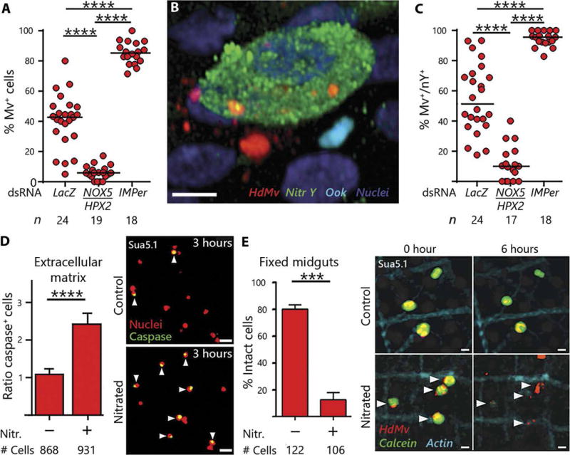
(A to C) Effect of reducing nitration by silencing NOX5/HPX2 or enhancing it by silencing IMPer on the proportion of invaded HdMv+ cells (A) and on the proportion of nitrated HdMv+ cells (C). Association of HdMv with nitrated cells undergoing apoptosis (B). HdMv, red; nitrotyrosine, green; ookinete, cyan; nuclei, blue. Medians are indicated by the black line and compared using the Mann-Whitney U test. (D) Effect of contact of Sua5.1 cells with a nitrated extracellular matrix on the rate of apoptosis, nuclei (red), and nuclei of caspase+ apoptotic cells (green; see arrowheads). (E) Effect of contact with nitrated midguts on cell integrity measured by retention of cytoplasmic calcein (green), HdMv (red), and actin (cyan). Line indicates medians that were compared using the Mann-Whitney U test (***P < 0.001, ****P < 0.0001). Scale bars, 10 μm.