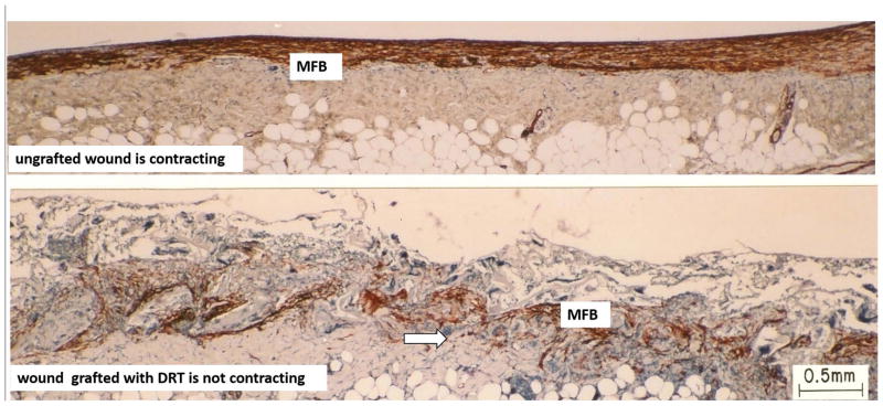
Figure 2. Skin wounds.
Sharply contrasting behavior during healing of two full-thickness skin wounds in the guinea pig. Histology sections were stained with antibody to α-smooth muscle actin. Top: Ungrafted wound is contracting vigorously on day 10. Dense assemblies of highly oriented contractile cells (myofibroblasts, MFB; red brown) populate the wound. Bottom: Wound grafted with a collagen scaffold, the dermis regeneration template (DRT), is not contracting on day 11. Grafting with DRT (bottom) resulted in significant reduction in MFB, dispersion of MFB assemblies and randomization of alignment of MFB axes. These changes describe a dramatic change in MFB phenotype and hypothetically account for the observed cancellation of the macroscopic contractile force in the wound. Red brown, MFB. Arrows: Scaffold struts. Scale bar: 0.5 mm. (adapted from (73)).

