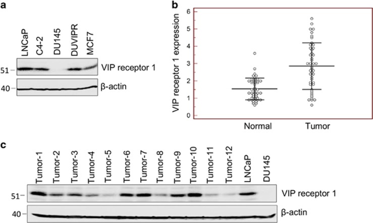
Figure 1.
Expression of VIP receptor in cell lines and tumors. (a) Cell lysates of indicated cells were western blotted, and the blot was developed with anti-VIPR1 antibodies. Equal loading was assessed by β-actin antibodies. (b) VIP receptor expression was measured by quantitative real-time PCR on malignant and non-malignant tumor adjacent normal tissue from breast cancer patients. In about 60% tumors VIPR1 expression was more than non-malignant tissue. (c) Protein was extracted from breast cancer tumors and 50 μg protein was western blotted, and the endogenous VIPR1 expression was detected

