Figure 3.
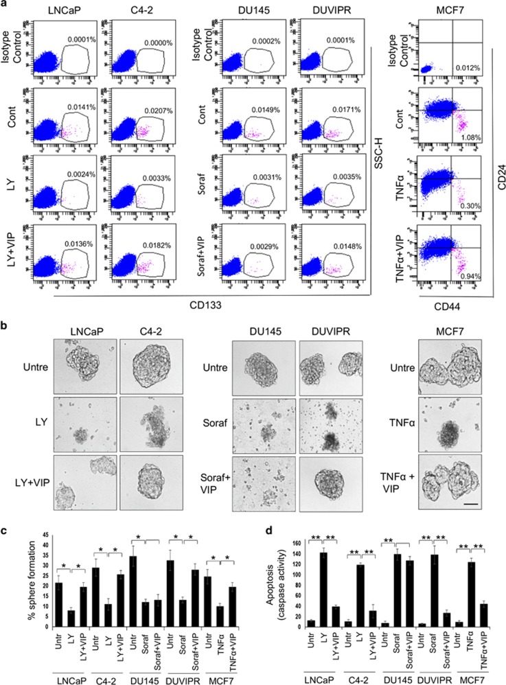
VIP protects spheres and purified CSCs from drug-induced apoptosis. (a) Indicated parental cells were starved for 12 h and apoptosis was induced by treating with either 20 μM LY, 20 μM sorafenib or 10 ng/ml TNFα. Fifteen minutes later, 100 nM of VIP was added. After 24 h, single cell suspensions were stained with CD133 antibodies, or double stained with CD44 and CD24 antibodies and analyzed by flow cytometry. Note that VIP restores the CSC population from drug-induced apoptosis. (b) Spheres derived from indicated cell lines were starved for 12 h and treated as in a. Phase-contrast images are shown. Scale bar=100 μm. (c) Spheres in b were dissociated, and sphere-forming assay was performed by culturing cells in sphere medium for 8 days on ultra-low attachment plates. The number of reformed spheres was counted. (d) Purified CSCs were treated as in a and the caspase activity in the cell lysates was measured using the fluorogenic substrate Ac-DEVD-AMC. The P-values for the indicated comparisons were obtained by two-tailed independent Student’s t-test. *P<0.05, **P<0.01

