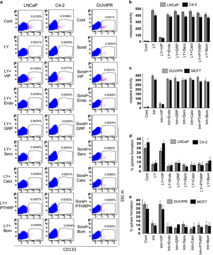
Figure 4.
Cytoprotective efficiency of various neuropeptides on CSCs. (a) Parental cells were treated as in Figure 3a to induce apoptosis. Fifteen minutes later, the cells were treated with 100 nM of each of indicated neuropeptide. Single cell suspensions were stained with antibodies against CD133 and analyzed by flow cytometry. (b and c) Spheres derived from indicated cell lines were starved for 12 h and treated as in a. Caspase activity in the sphere cell lysates was measured as in Figure 3d. Inhibitor=Sorafenib for DUVIPR cells, and TNFα for MCF7 cells. (d and e) Spheres in b and c were dissociated, and sphere-forming assay was performed, and the number of reformed spheres was counted. Results suggest that antiapoptotic effect is specifically exhibited by VIP. Please see Supplementary Table 1 for P-values

