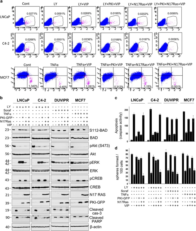
Figure 7.
VIP-induced cytoprotection is abrogated by dominant-negative PKI-GFP and N17 Ras. (a) Parental cells expressing either empty vector or PKI-GFP and/or HA-N17 Ras were treated as in Figure 6a. Individual cells were stained with antibodies against CD133 or CD44/CD24 and analyzed by flow cytometry. (b) Purified CSCs, expressing PKI-GFP and/or HA-N17 RAS were treated with indicated inhibitors. VIP was added 15 min later. BAD phosphorylation was detected as in Figure 5a. Blots of S112-BAD, phospho-Akt, phospho-ERK and phospho-CREB were, respectively, stripped and reprobed with total BAD, Akt, ERK and CREB. Expression of PKI-GFP and HA-N17 Ras were, respectively, detected using antibodies against GFP and HA. Cleavage products of PARP and caspase-3 were used as apoptotic markers in whole-cell lysates. (c) Caspase activity in cell lysates of b was measured as described in Figure 3d. (d) Sphere-forming assay was performed using cells in b, and the number of spheres generated was counted. The error bars represent S.D. of the biologic triplicates. All experiments presented in this figure are representative of two independent experiments. Please see Supplementary Table 3 for P-values

