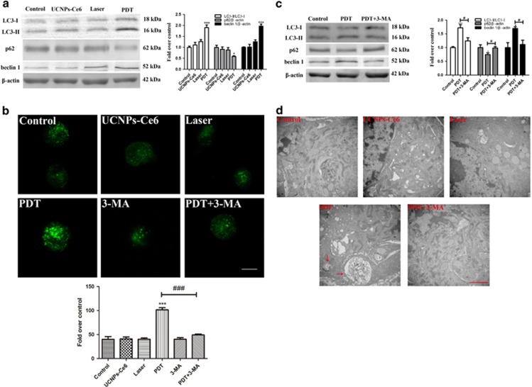
Figure 3.
Autophagy induced by PDT is dependent on the combined effects of the laser and UCNPs-Ce6. (a) The expression levels of the autophagy-related proteins LC3, p62 and beclin 1 2 h after the various treatments. (b) The observation of AVOs induced by various treatments using MDC staining (scale bar, 5 μm). (c) The effects of 3-MA on the expression levels of the autophagy-related proteins LC3, p62 and beclin 1 2 h after PDT. (d) Morphological alterations of THP-1 macrophage-derived foam cells following various treatments detected via representative transmission electron microscopy (red arrow: autophagic vacuoles with cytoplasmic contents, scale bar, 2 μm); (n=3; *P<0.05, ***P<0.001 versus control group, ###P<0.001 versus PDT group)

