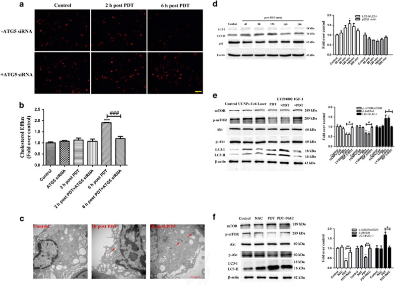
Figure 7.
Photodynamic effects on peritoneal macrophage-derived foam cells. (a) The effect of PDT on the intracellular lipid burden measured by Dil ox-LDL staining (scale bar, 50 μm). (b) Quantitative detection of the cholesterol efflux following PDT using a fluorometric assay. (c) Morphological alterations following PDT detected via representative transmission electron microscopy (red arrow: autophagic vacuoles with cytoplasmic contents, scale bar, 2 μm). (d) The expression levels of the autophagy-related proteins p62 and LC3 from 40 min to 200 min post-PDT. (e) The effects of LY294002 and IGF-1 on the expression levels of PI3K/Akt/mTOR pathway-related proteins and the autophagy-related protein LC3 2 h post-PDT. (f) The effects of NAC on the expression levels of PI3K/Akt/mTOR pathway-related proteins 2 h post-PDT (n=3; *P<0.05, **P<0.01, ***P<0.001 versus control group, # P<0.05, ##P<0.01, ###P<0.001 versus PDT group)

