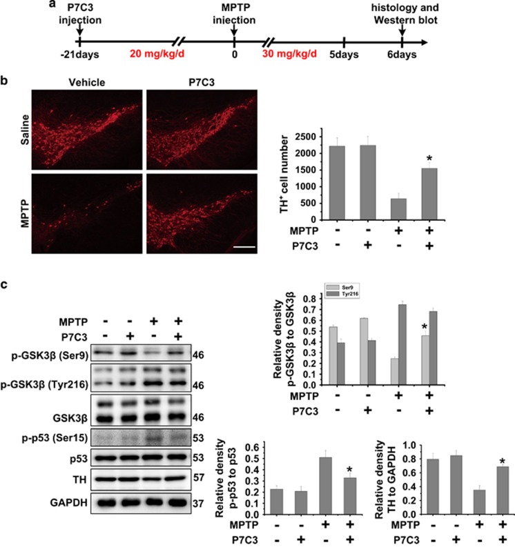
Figure 6.
P7C3 inhibits GSK3β activity to protect DA neurons from MPTP toxicity in vivo. (a) A schematic diagram of animal experimental procedure is shown. (b) Twenty-four hours after the last intraperitoneal injection, mice were killed and fixed with perfusion. The brains were post-fixed with a fixing agent overnight at 4 °C, followed by the treatment with 30% sucrose at 4 °C for another night. Serial 20 μM-thick mouse midbrain slices were cut using a freezing microtome. Immunohistochemical staining was conducted using anti-TH antibody. The quantification of TH+ cell numbers is shown in the right panel. Scale bar, 10 μm. n=4 per group. (c) Twenty-four hours after the last intraperitoneal injection, the fresh midbrains samples were collected and subjected to immunoblot analysis with indicated antibodies. n=4 per group. The protein levels of p-GSK3β (Ser9), p-GSK3β (Tyr216), GSK3β, p53, p-p53, TH and GAPDH were measured using immunoblot analysis. The quantification of the intensity of p-GSK3β (Ser9) and p-GSK3β (Tyr216) relative to GSK3β, p-p53 relative to p53, and TH relative to GAPDH are shown in the right panels. n=4 per group. The values are presented as means ± S.E.M. from three independent experiments. *P<0.05 versus the group treated with MPTP alone using one-way ANOVA

